Anatomy & Physiology
Learn the structure and function of the human body systems, from cells to organs.
Nervous System
Sign in to track progress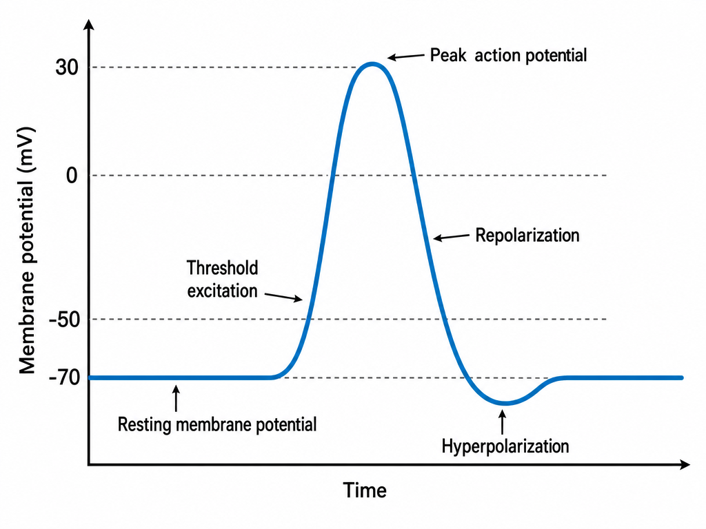
The nervous system is the body's control center, coordinating sensation, movement, thought, and homeostasis.
Divisions of the Nervous System
- Central Nervous System (CNS): Brain and spinal cord
- Peripheral Nervous System (PNS): All nerves outside the CNS
- Somatic: Voluntary control of skeletal muscle
- Sensory input
- Motor input
- Autonomic: Involuntary control of organs and glands
- Sympathetic: "Fight or flight" response
- Parasympathetic: "Rest and digest" response
- Somatic: Voluntary control of skeletal muscle
Neuron Structure
Neurons consist of:
- Cell body (soma): Contains nucleus and organelles
- Dendrites: Receive signals from other neurons
- Axon: Transmits signals away
- Synapse: Junction between two neurons
Neurotransmission
- Action potential arrives at axon terminal
- Voltage-gated Ca²⁺ channels open
- Ca²⁺ influx triggers vesicle fusion
- Neurotransmitter is released into synaptic cleft
- Neurotransmitter binds to receptors on postsynaptic membrane
- Signal is propagated or inhibited
- Neurotransmitter is removed by reuptake, enzymatic breakdown, or diffusion
Neuromuscular Junction The neurotransmitter released at the neuromuscular junction is acetylcholine (ACh). Acetylcholine binds to nicotinic receptors on the muscle membrane, leading to depolarization and muscle contraction.
Blood-Brain Barrier (BBB)
The BBB is a selective barrier formed by tight junctions between capillary endothelial cells. It protects the brain by restricting which substances can enter from the blood; lipid-soluble, small, or uncharged molecules cross more easily than large or charged ones.
Action Potentials & the All-or-None Principle
An action potential is a rapid depolarization of a neuron membrane driven by voltage-gated Na⁺ and K⁺ channels. It is all-or-none: once threshold is reached, the action potential fires at full amplitude; subthreshold stimuli produce no action potential. This ensures reliable signal transmission over long distances.
Action Potential Phases
Neuronal action potentials move through distinct phases driven by voltage-gated ion channels:
- Resting potential: The membrane stays polarized at rest, maintained mainly by K⁺ leak channels and the Na⁺/K⁺ ATPase.
- Depolarization: Voltage-gated Na⁺ channels open, allowing Na⁺ influx and making the inside of the cell more positive.
- Repolarization: Na⁺ channels inactivate and voltage-gated K⁺ channels open, allowing K⁺ efflux that restores the negative resting membrane potential.
- Hyperpolarization: K⁺ channels remain open briefly, causing the membrane potential to dip below resting before returning to baseline.
Brain Regions
- Cerebrum: Largest region, upper part of the brain. Controls higher brain functions, including thinking, memory, learning, language, sensory processing, emotions, and voluntary movement.
- Cerebellum: Below the cerebrum, at the back of the brain. Coordinates balance, posture, muscle coordination, and fine motor movements.
- Brainstem: Base of the brain, connecting the brain to the spinal cord. Regulates automatic functions such as breathing, heart rate, blood pressure, swallowing, and consciousness. It contains three parts:
- Midbrain: Controls eye movements, visual and auditory reflexes, and motor control.
- Pons: Acts as a bridge between the cerebrum and cerebellum; helps regulate breathing, sleep, and facial movements.
- Medulla Oblongata: Regulates vital functions including breathing, heart rate, blood pressure, swallowing, coughing, sneezing, and vomiting.
- Hypothalamus: Below the thalamus, near the center/base of the brain. Maintains homeostasis by regulating body temperature, hunger, thirst, sleep, hormone release (via the pituitary gland), and autonomic nervous system functions.
- Thalamus: Deep in the center of the brain, above the brainstem. Acts as the brain's sensory relay station, sending most sensory information to the cerebral cortex, and it regulates alertness and consciousness.
Cerebral Cortex
The cerebral cortex is the outermost layer of the brain. It has many folds that increase its surface area and allow it to contain more neurons.
The cerebral cortex is divided into four main lobes:
1. FRONTAL LOBE
- Location: Front of the brain
- Main roles:
- Planning
- Decision-making
- Problem-solving
- Voluntary movement
- Speech production
- Behavior and personality
- Prefrontal Cortex: Controls planning, judgment, attention, problem-solving, and self-control.
- Motor Cortex: Controls voluntary body movements, such as moving the arms, legs, hands, and face.
- Broca's Area: Helps produce speech.
2. PARIETAL LOBE
- Location: Upper and back portion of the brain
- Main roles:
- Touch
- Pressure
- Pain
- Temperature
- Body awareness
- Spatial awareness
- Somatosensory Cortex: Receives sensory information from the body, including touch, pain, pressure, and temperature.
3. OCCIPITAL LOBE
- Location: Back of the brain
- Main role: Vision
- The occipital lobe receives and processes visual information from the eyes and helps the brain recognize shapes, colors, movement, and objects.
4. TEMPORAL LOBE
- Location: Lower sides of the brain, near the ears
- Main roles:
- Hearing
- Sound processing
- Language comprehension
- Memory
- Auditory Cortex: Processes sounds received from the ears.
- Wernicke's Area: Helps understand spoken and written language.
Key Neurotransmitters
- Acetylcholine (ACh):
- Major neurotransmitter of the parasympathetic nervous system
- Released by all autonomic preganglionic neurons
- Released at the neuromuscular junction to stimulate skeletal muscle contraction
- Nicotinic receptors: ligand-gated ion channels
- Muscarinic receptors: G protein-coupled receptors (GPCRs)
- Norepinephrine (NE):
- Major neurotransmitter released by most sympathetic postganglionic neurons
- Acts primarily at α₁, α₂, and β₁ receptors, with less activity at β₂ receptors
- Dopamine:
- Important in movement, motivation, reward, and cognition
- Acts at D₁–D₅ receptors
- Dopamine receptors are GPCRs
- Serotonin (5-HT):
- Involved in mood, sleep, appetite, nausea, and gastrointestinal function
- Has multiple receptor families and subtypes (e.g., 5-HT₁A, 5-HT₂A)
- GABA (γ-Aminobutyric Acid):
- Major inhibitory neurotransmitter in the central nervous system
- Generally decreases neuronal excitability
- Glutamate:
- Major excitatory neurotransmitter in the central nervous system
- Important in learning, memory, and synaptic plasticity
Key Points
- The autonomic nervous system has sympathetic and parasympathetic divisions
- Neurotransmission involves neurotransmitter release, receptor binding, and removal
- Major neurotransmitter systems include glutamate, GABA, dopamine, serotonin, norepinephrine, and acetylcholine
- The blood-brain barrier selectively restricts substances entering the CNS from the blood
- Lipid-soluble and small molecules cross the BBB more easily
- Action potentials are all-or-none depolarizations driven by Na⁺ and K⁺ channels
- Neuronal action potentials progress through resting, depolarization, repolarization, and hyperpolarization phases
- Major brain regions include the cerebrum, cerebellum, brainstem, hypothalamus, and thalamus
- The cerebral cortex has four main lobes: frontal, parietal, occipital, and temporal
- The frontal lobe controls planning, decision-making, voluntary movement, and speech production
- The parietal lobe processes touch, pain, temperature, and spatial awareness
- The occipital lobe processes visual information such as shapes, colors, and movement
- The temporal lobe handles hearing, language comprehension, and memory